How to Read a Peptide LC–MS Result: m/z, Charge and Intact Mass
Check ion charge, adducts and mass error, then judge what the evidence supports about peptide identity.
Compare the observed m/z with the expected ion for the same charge and adduct, or compare deconvoluted and theoretical masses on the same mass basis. A compatible intact mass supports identity. Sequence and stereochemistry may require additional evidence.
Read the chromatogram and spectrum together
Liquid chromatography separates components over time. The mass spectrum shows ion signals by mass-to-charge ratio, written m/z. First identify the chromatographic peak or retention interval from which the spectrum was taken. A total ion current chromatogram sums the measured ion signal at each time point across the mass range used, while an extracted ion chromatogram follows a selected m/z window. A strong extracted trace tells you when ions in that window appeared. Other ions can appear at the same time.
A spectrum can contain protonated ions, adducts and isotope peaks. Check the assigned charge and ion formula before comparing a peak with a theoretical value. Also check whether the report gives the mass of the monoisotopic species, calculated from specific isotope masses, or an average mass based on isotope abundances. A deconvoluted neutral mass is derived from the measured ion signals and their assignments.
IUPAC, Definitions of terms relating to mass spectrometry (2013)
Work through charge and mass error
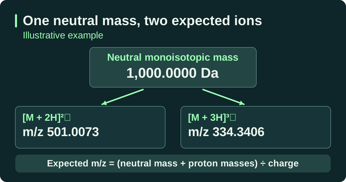
The worked example uses a neutral monoisotopic model mass of 1,000.0000 Da. For a protonated ion, its expected m/z is the sum of the neutral mass and the added proton masses divided by the charge. When you calculate a signed error in ppm, compare observed and expected values on the same basis. Use the expected m/z for an ion comparison or the expected neutral mass for a comparison after deconvolution.
Suppose the doubly charged ion was measured at m/z 501.0088. With unrounded theoretical values, the signed mass error is about +3.0 parts per million. The calculation is one million times the difference between observed and expected m/z, divided by expected m/z. When a report compares neutral masses instead, use the expected neutral mass as the denominator and keep that basis in the record. Evaluate the resulting difference against the method’s stated tolerance for that measurement.
For these fictional monoisotopic ions, each positive charge comes from a proton. With proton mass mp ≈ 1.007276 Da and charge z, the neutral mass is z × (m/z − mp). Use the assigned ion formula if the report indicates another adduct.
| Assigned ion | Charge | Expected m/z, rounded | Reverse calculation using rounded values | Neutral mass |
|---|---|---|---|---|
| [M + 2H]²⁺ | +2 | 501.0073 | 2 × (501.0073 − 1.007276) | ≈1,000.000 Da |
| [M + 3H]³⁺ | +3 | 334.3406 | 3 × (334.3406 − 1.007276) | ≈1,000.000 Da |
In a separate neutral-mass comparison, a deconvoluted result of 1,000.0030 Da against 1,000.0000 Da gives +3.0 ppm using (observed − expected) ÷ expected × 1,000,000. The fictional report MS-DEMO-02 specifies ±5 ppm for neutral monoisotopic mass, so the mass agrees within that report’s tolerance.
Intact-mass finding. The result supports the expected intact mass for the reported sample. Sequence evidence. The report supplies no fragment assignments, so residue order remains unresolved. For a decision about structural identity, check the evidence for the method’s intended use.
IUPAC, Definitions of terms relating to mass spectrometry (2013) · NIST, 2022 CODATA recommended values, proton mass in u
Distinguish evidence for mass, sequence and configuration
Different molecules can share the same intact mass. Leucine and isoleucine have the same elemental composition, and replacing an L residue with its D counterpart preserves mass. For sequence evidence, look for fragment ions in an MS/MS report and ask which part of the sequence the assigned fragments cover. Ordinary mass matches may leave isobaric residues or stereochemistry unresolved. Depending on the ambiguity, a suitable reference comparison, stereochemical analysis or another complementary procedure may be needed.
The original reference-standard study by McCarthy and colleagues describes using multiple techniques to characterize peptide standards, including mass spectrometry and other tests for structural features that may need additional evidence. EMA’s synthetic peptide guideline discusses complementary identity tests in the context of medicinal product applications.
A usable interpretation can be as short as “At 5.21 min the observed [M + 2H]²⁺ ion is consistent with the expected monoisotopic m/z within this method’s tolerance. The report provides no fragment assignments.” Keep the spectrum, retention window, charge/adduct assignment and theoretical calculation with the note.
McCarthy et al. (2023), Reference Standards to Support Quality of Synthetic Peptide Therapeutics · EMA, Synthetic peptides guideline, §§ 2, 4.1, 4.3 and 4.4